We found that HEK293T EV uptake is an active process that is dose and time dependent. Here we optimized an imaging flow cytometry IFC-based platform to quantitatively assess dose time and recipient cell specificity effects on human embryonic kidney cell HEK293T EV internalization in a high-throughput manner.

Improved Loading Of Plasma Derived Extracellular Vesicles To Encapsulate Antitumor Mirnas Molecular Therapy Methods Clinical Development
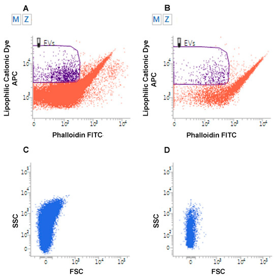
EV-associated markers were analyzed by flow cytometry CytoFLEX S Beckman Coulter USA.

. By staining different cargo components we were able to directly. By labeling EVs with PKH26 or PKH67 the interactions with and uptake of EVs by epithelial and breast cancer cells 13 macrophages 43 dendritic cells 29 and endothelial and myocardial cells 44 have been followed using fluorescence microscopy and flow cytometry. 12Ellison Brian J.
The characterization of EV uptake notably specificity dose and time dependence and kinetic assays will help inform and develop targeted and efficient EV-based therapeutics. Confocal imaging and flow cytometry analysis of EV uptake To complement the SPR data with other independent and commonly used methods the uptake of PLT and RBC EVs by PC-3 cells was investigated by confocal fluorescent imaging and flow cytometry. In vitro EV imaging helps researchers to understand the physical property of EVs such as the mechanism of EV release and uptake 1 29 or biomarkers expressed on the EV surface 30 31.
Here we optimized an imaging flow cytometry IFC-based platform to quantitatively assess dose time and recipient cell specificity effects on human embryonic kidney cell HEK293T EV internalization in a high-throughput manner. Both sEVs and mEVs secreted by HEK293T-palmGFP cells were bound onto the surface of latex beads 3 µm 4 wv AldehydeSulfate Latex Beads ThermoFisher USA overnight at 4 C. The lipid content of EVs was used as a basis of normalization in our EV uptake studies.
Extracellular vesicles EVs exosomes microvesicles microparticles vesicles 37 imaging flow cytometry IFCM analyses of EVs lipid dye 38 Introduction. 103389fimmu201801011 Monitoring extracellular Vesicle Cargo Active Uptake by Imaging Flow Cytometry Yifat. PDF Abstract Extracellular vesicles EVs are nanosized lipid bilayer-bound vesicles that are naturally secreted from most cell types as a.
Vesicle Cargo Active Uptake by Imaging Flow Cytometry. 35 EV characterization tools which are able to localize and confirm specificity of EV labelling. Flow cytometry of EVs.
2B and Supplementary Fig. Here we present an imaging flow cytometry IFC method for monitoring the uptake of malaria-derived vesicles by host immune cells. Independent of EV source MDA-MB-231 cells consistently exhibited more EV uptake than did MCF-7 cells and HEK293FT cells exhibited no observable EV uptake suggesting that recipient cell type is the major factor in determining EV uptake among these cases Fig.
Relative amounts of EVs 100 50 10 and 0 were stained using the. Utilizing an imaging flow cytometry platform IFC HEK293T EV uptake was analyzed. Whereas uptake of EVs has been amply demonstrated by live imaging visualization of EV interaction with the PM has been reported on just a few occasions 60 and only recently with live imaging.
EV uptake by cells can be visualized directly by use of fluorescent lipid membrane dyes that stain the EV membranes. Although uptake of HEK293FT-derived EVs appeared higher across recipient. We found that HEK293T EV uptake is an active process that is dose and time dependent.
By combining recent advancements in imaging flow cytometry for the phenotypic analysis of single EVs and aiming to identify target cells for EVs within therapeutically relevant MSC-EV preparations we explored the EV labelling efficacy of various fluorescent dyes specifically of CFDA-SE Calcein AM PKH67 BODIPY-TR-Ceramide and a novel lipid dye named Exoria. Pf-derived EVs were labeled. Here we optimized an imaging flow cytometry IFC-based platform to quantitatively assess dose time and recipient cell specificity effects on human embryonic kidney cell HEK293T EV internalization in a high-throughput manner.
HEK293T EV uptake was dose and time dependent with a minimum threshold dose of 6000 EVs per cell at 4. ImageStream Acquisition was performed on the ImageStreamX Mark II Imaging Flow Cytometer Luminex Corporation Seattle Washington using the INSPIRE software. In vivo EV imaging aids in unveiling the biodistribution of EVs which can be used to characterize pharmacokinetic property of EVs as a drug andor theranostics vehicle.
Further the selectivity of EV uptake was quantified in vitro and we found. However imaging and tracking. Uptake Using Imaging Flow Cytometry Taylor J.
A minimum of 500010000 cell events were acquired. Download scientific diagram Relative Plasmodium falciparum Pf-derived extracellular vesicles EV uptake into monocytes. We found that HEK293T EV uptake is an active process that is dose and time dependent.
Find read and cite all the research you need on. Extracellular vesicles Selective uptake HEK293T Imaging flow cytometry Introduction Extracellular vesicle research is a burgeoning field due to the therapeutic and diagnostic utility of natural and. Download scientific diagram Kinetics measurement of Plasmodium falciparum Pf-extracellular vesicles EV uptake into monocytes using imaging flow cytometry IFC.
For both cell culture and in vitro EV uptake platforms 7. Further the selectivity of EV uptake was quantified in vitro and we found.
Ijms Free Full Text Extracellular Vesicles As Signaling Mediators And Disease Biomarkers Across Biological Barriers Html
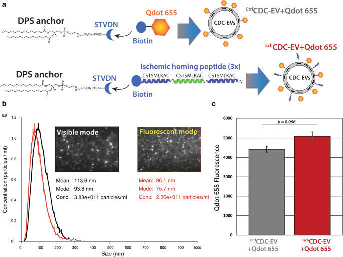
Targeting Extracellular Vesicles To Injured Tissue Using Membrane Cloaking And Surface Display Springerlink
![]()
Pdf Routes And Mechanisms Of Extracellular Vesicle Uptake

Technologies And Standardization In Research On Extracellular Vesicles Trends In Biotechnology

Mechanisms And Modulation Of Microvesicle Uptake In A Model Of Alveolar Cell Communication Journal Of Biological Chemistry
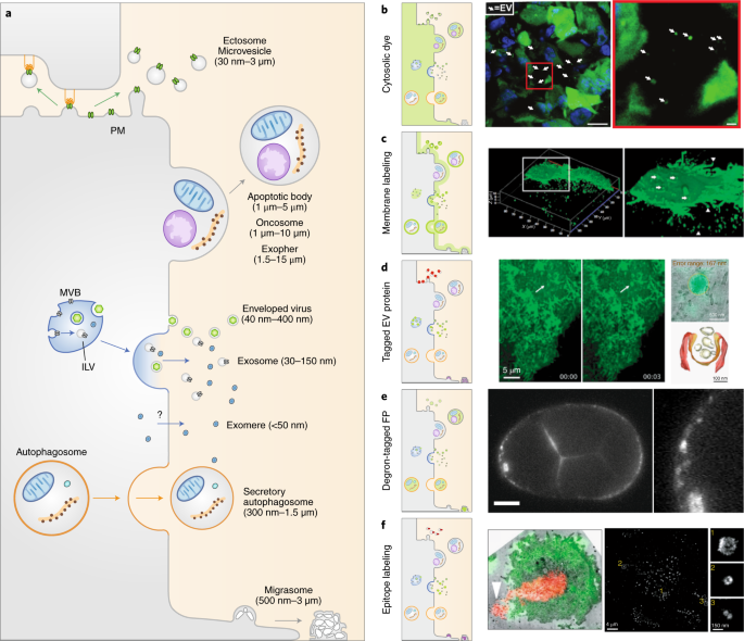
The Power Of Imaging To Understand Extracellular Vesicle Biology In Vivo Nature Methods

Molecular Evaluation Of Five Different Isolation Methods For Extracellular Vesicles Reveals Different Clinical Applicability And Subcellular Origin Veerman 2021 Journal Of Extracellular Vesicles Wiley Online Library

Biological Membranes In Ev Biogenesis Stability Uptake And Cargo Transfer An Isev Position Paper Arising From The Isev Membranes And Evs Workshop Russell 2019 Journal Of Extracellular Vesicles Wiley Online Library
Technologies And Standardization In Research On Extracellular Vesicles Trends In Biotechnology

Dir Labelling Does Not Affect Ev Morphology And Co Localizes With Evs Download Scientific Diagram

Mechanisms And Modulation Of Microvesicle Uptake In A Model Of Alveolar Cell Communication Journal Of Biological Chemistry
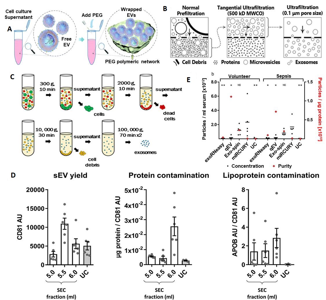
Isolation And Analysis Methods Of Extracellular Vesicles Evs
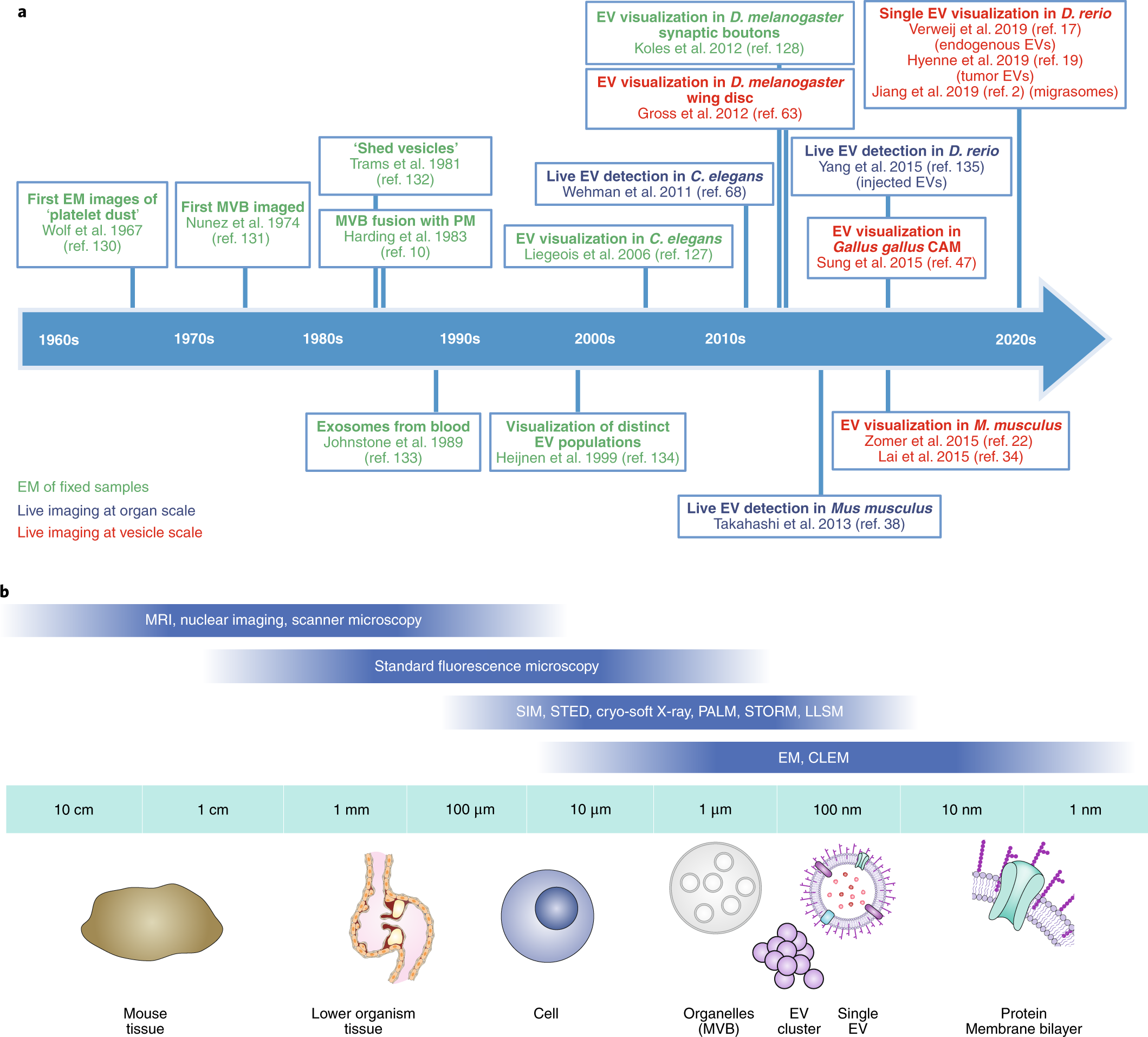
The Power Of Imaging To Understand Extracellular Vesicle Biology In Vivo Nature Methods
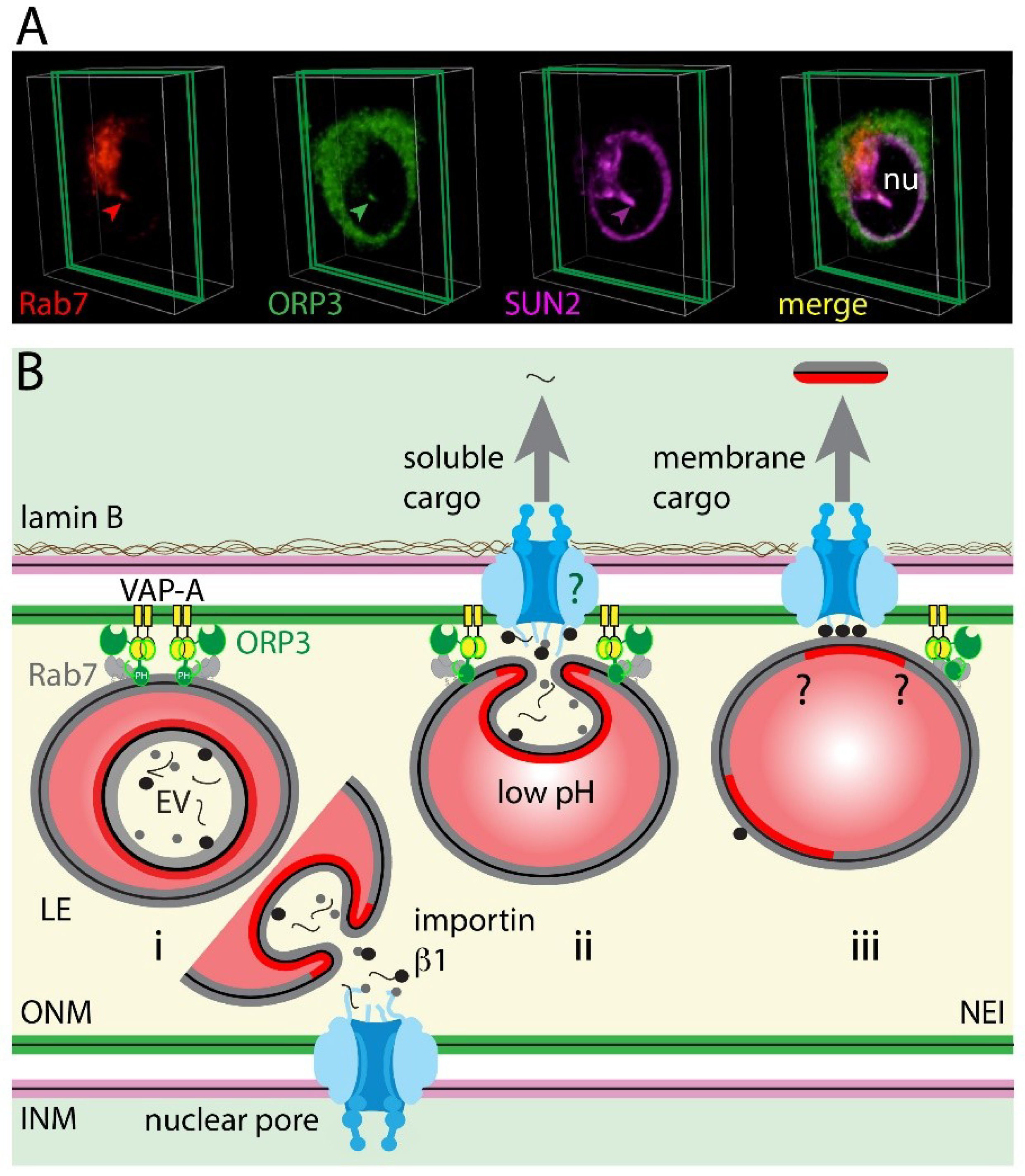
Cells Free Full Text Uptake And Fate Of Extracellular Membrane Vesicles Nucleoplasmic Reticulum Associated Late Endosomes As A New Gate To Intercellular Communication Html

Improved Loading Of Plasma Derived Extracellular Vesicles To Encapsulate Antitumor Mirnas Molecular Therapy Methods Clinical Development

Dir Labelling Does Not Affect Ev Morphology And Co Localizes With Evs Download Scientific Diagram

Technologies And Standardization In Research On Extracellular Vesicles Trends In Biotechnology
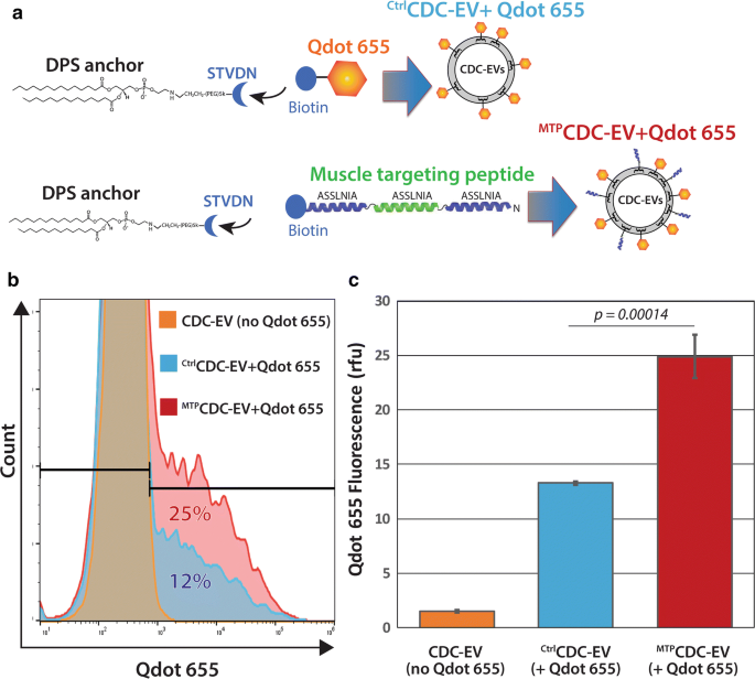
Targeting Extracellular Vesicles To Injured Tissue Using Membrane Cloaking And Surface Display Springerlink

Quantification Of Extracellular Vesicles In Vitro And In Vivo Using Sensitive Bioluminescence Imaging Gupta 2020 Journal Of Extracellular Vesicles Wiley Online Library


0 komentar:
Posting Komentar